DVT – Phlebothrombosis
One of the serious complications of being immobile and bedridden for a longer period is deep vein thrombosis (DVT), as it can cause pulmonary embolism and death.
What is DVT?
From the name, we can get some idea that it is a thrombus in the deep veins. This thrombus formation is due to various causes, all of which come under Virchow’s triad.
Virchow’s Triad
- Venous stasis
- Vein wall injury
- Hypercoagulable state
Favourable sites for DVT
Deep vein thrombosis is common in leg veins - soleal veins, femoral veins, popliteal veins, and pelvic veins.
Upper limb veins such as axillary veins are also affected in certain conditions causing compression of veins.
People at Risk
- Patients who are bedridden for a longer period.
- Postoperative immobilization for more than 3 days
- Long flight travel - Traveller’s thrombosis
- Pregnancy - due to the compression of inferior vena cava by the gravid uterus.
- Malignant state of the pancreas, the stomach can also produce deep vein thrombosis
- Women using oral contraceptive pills
- Smoking, as it increases the viscosity of the blood, thereby facilitating thrombus formation.
- Polycythemia vera, thrombocytosis
- Homocysteinemia - damage to the collagen in the blood vessel wall.
- Nephrotic syndrome - loss of antithrombin in the urine
All these conditions make the person prone to develop deep vein thrombosis in the leg veins.
Some other conditions leading to DVT in axillary veins are
- Repeated central venous catheterization.
- Cervical rib
- Thoracic inlet syndrome
- Radiotherapy to axilla
- Axillary lymph node block dissection
- People who use their arms for a long time working such as swimming, football, wrestling, etc.
Wisdom: In these people, there can be hypertrophy of the scalene muscle resulting in compression of veins. This condition is given a special name called Paget-Schroetter syndrome.

Genetic factors such as hereditary thrombosis syndrome, deficiency of protein C or S can also make the people more prone to develop deep vein thrombosis.
The Patient
Patient in acute condition will present with
- Swelling of the affected part - pitting edema
- Warmth and tenderness over the affected part
- Dorsiflexion of foot produce pain over the calf - positive Homan’s sign
- History of recent travel

Patient in chronic condition will present with
- Varicose veins, which are abnormally dilated and tortuous veins.
- Skin ulcerations due to venous stasis
- A brownish discoloration of the skin due to deposition of hemosiderin from the breakdown of RBCs present in the stasis blood
A patient who developed pulmonary embolism will present with
- Hemoptysis
- Breathlessness
- Chest pain
Differential Diagnosis
Some diseases have similar features of DVT. They are:
Ruptured baker's cyst - swelling will occupy behind the knees and there will be predisposing conditions like rheumatoid arthritis.
Cellulitis and superficial thrombophlebitis can mimic DVT.
Pitting edema could be caused by cardiac failure, hypoproteinemia, or nephrotic syndrome.
Sequelae
DVT may dislodge to form pulmonary embolism, form venous gangrene, cause dermatitis, or lead to acute bleeding as a result of treatment with anticoagulant drugs.
Work Up
Venous duplex ultrasonography is very sensitive and specific for diagnosing deep vein thrombosis.
The D-dimer level can help us to rule out DVT. It is generally elevated in active lysis of the thrombus. Normal level is < 500mg/ml.
The coagulation profile of the patient has to be checked. CT pulmonary angiogram is done for identifying pulmonary embolism.
Management
Treatment is by medical approach and preventing pulmonary embolism before it develops.
It is done by grading the patients as per modified Well’s criteria and treating them accordingly. It includes components like presence of tenderness, swelling of the limb, cancer history, history of being bedridden, any operation history, etc.
- Low-risk patients have score 0,
- Moderate-risk patients are ones with score 1 to 2 and
- High-risk patients have scores above 2.
For fixed thrombus, patients are given low dose molecular weight heparin. If they develop thrombocytopenia they are switched to fondaparinux. Recently approved drugs such as Rivaroxaban, Dabigatran, Apixaban, etc., can also be used.
For free thrombus, fibrinolytic agents such as streptokinase are used.
IVC filters such as Kim ray, Greenfield filter, Mobin-Uddin umbrella filter are used in patients not responding to medical treatment.
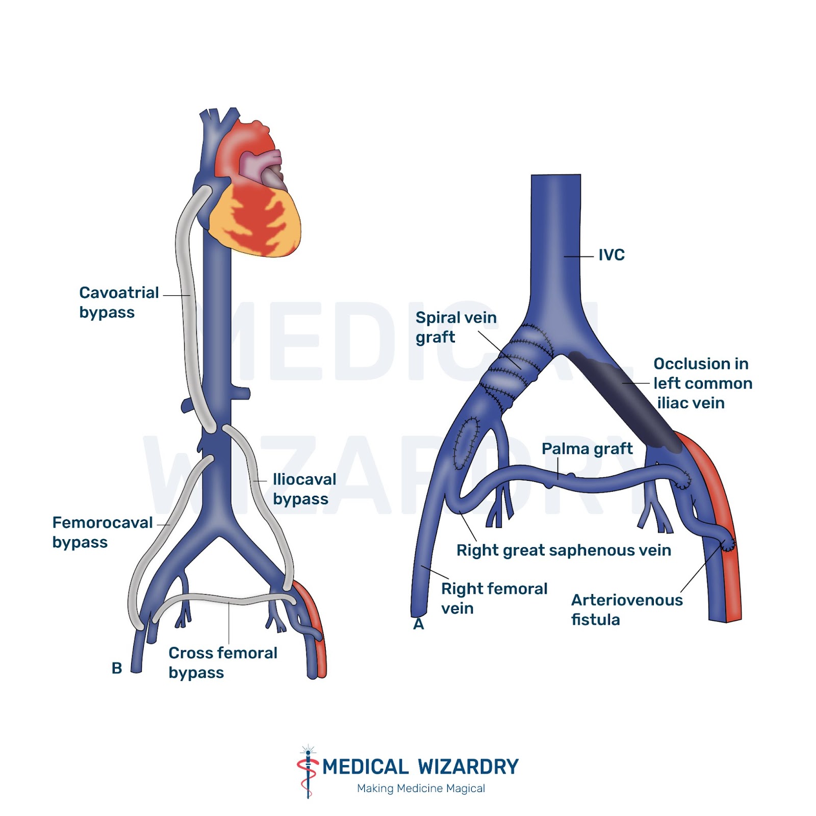
Operative procedures such as Palma operation for iliofemoral thrombosis and May-Husni operation for the thrombus in the popliteal vein are also practiced.
Preventing DVT
Smoking has to be avoided. Pressure bandage can be given or massaging the limbs can be done after major surgery.Various measures like pneumatic compression, electrical stimulation of calf muscles can be done to prevent the sluggish flow of blood through the vessels.
References
- SRB Manual of Surgery 6th ed; Page no: 216 - 219
- Bailey & Love’s Short practice of Surgery, 26th ed; Page no: 278,916.
- https://www.ncbi.nlm.nih.gov/books/NBK507708/
- https://www.ncbi.nlm.nih.gov/pmc/articles/PMC5778510/
- https://www.ncbi.nlm.nih.gov/books/NBK482420/
Author’s Footnote
DVT is characterized by the presence of the following signs:
- Homan’s sign - calf tenderness on forced ankle dorsiflexion
- Phlegmasia alba dolens - painful white leg
- Phlegmasia cerulea dolens - painful blue leg
- Moses sign - calf tenderness on direct pressure
- Pratt’s sign - calf tenderness on squeezing from the sides
Feel free to click on the references for a more in-depth reading if you so desire. If you feel any information can be added or that there are any inadvertent errors, please let us know in the comments below or bring it to our notice via support@medicalwizardry.com.
Ever otherwise, feel free to use the comments section for discussion.
We would love to hear from you. Reach out to us via admin@medicalwizardry.com to share any random gibberish/ideas that you would like implemented.