From making our muscles move to storing our deepest memories, neurons play a fascinating role as the building blocks of the nervous system.
To understand how microscopic neurons accomplish these seemingly impossible tasks, we need to understand nerve conduction.
It is essentially a mechanism by which messages are transmitted across the body. To put in simple words, it’s the way the nerves talk to one another.
As always, let’s review a bit of the structure of the nerve before going into the details of nerve conduction.
Take a close look at the image provided below:

Author: DrV
The nerve cell consists of a cell body (also called cyton) from which short thread-like structures project outward. These are called dendrites.
The cyton also projects into a longer thread-like structure - the axon. The axon is covered by multiple Schwann cells, which form a myelin sheath.
The myelin sheath is not a continuous structure. It has gaps called nodes of Ranvier.
Each axon terminates by branching out into axonal terminals.
So when you think of nerve conduction, the first thing that pops in your head is the synapse. The synapse is the space between the knob of the first neuron and the dendrite of the second. This is where the neurotransmitters are “poured” into during nerve conduction.
Role of Myelin
The myelin sheath of a neuron is similar to the insulation of an electric wire. The part of the neuron that is covered by myelin does not conduct electricity.
Myelination in the peripheral nervous system is carried out by Schwann cells. Each peripheral nerve is wrapped around by multiple Schwann cells.
However, the story is different in the case of the central nervous system, where the neurons are myelinated by the oligodendrocytes. Each oligodendrocyte myelinates multiple neurons simultaneously, as shown below:

Author: Holly Fischer
Myelin allows the action potential (the impulse conducted) to jump from one node of Ranvier to the next without passing through the entire length of the axon.
This type of conduction is called saltatory conduction. Take a look at the image below:

Without myelin, the impulse will have to travel the entire length of the axon, thus making the conduction slow.
Did you know that myelination can increase the velocity of conduction more than 10 - 15 times?! Unmyelinated neurons have a conduction velocity of about 0.5 - 10 m/s, while myelinated neurons can have a conduction velocity of up to 150 m/s!
Nerve Conduction
A feature of neurons is their ability to conduct impulses. To understand this phenomenon, we need to first understand what resting membrane potential means.
A potential difference is said to exist when there is a difference in the level of something (for example, electric charge) across a clear boundary. In a cell, this potential difference is due to the unequal distribution of ions.
Normally, there is a large amount of K+ ions inside than outside the cell. Conversely, there’s a large number of Na+ ions outside the cell than within.
This balance is maintained by Na-K-ATPase. Such a gradient allows the passive diffusion of K+ outside the cell and Na+ into it (that is, the ions move along the gradient without spending ATP.
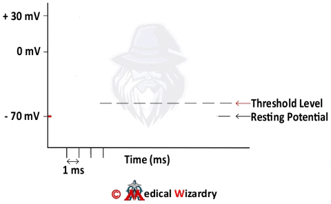
At rest: The membrane potential of a neuron is -70mV. In the unexcited state, the membrane is more permeable to K+. (This is because there are more open K+ channels at -70 mV as the equilibrium potential of K+ is close to -70 mV).

The Action Potential
Once the neuron receives a stimulus, the Na+ channels open and Na+ ions rush into the cell and raise the potential of the neuron to the threshold potential. This is depolarisation.
This immediately causes a positive feedback loop. In this loop, the opening of Na+ channels and the movement of Na+ into the cell only causes more Na+ channels to open.
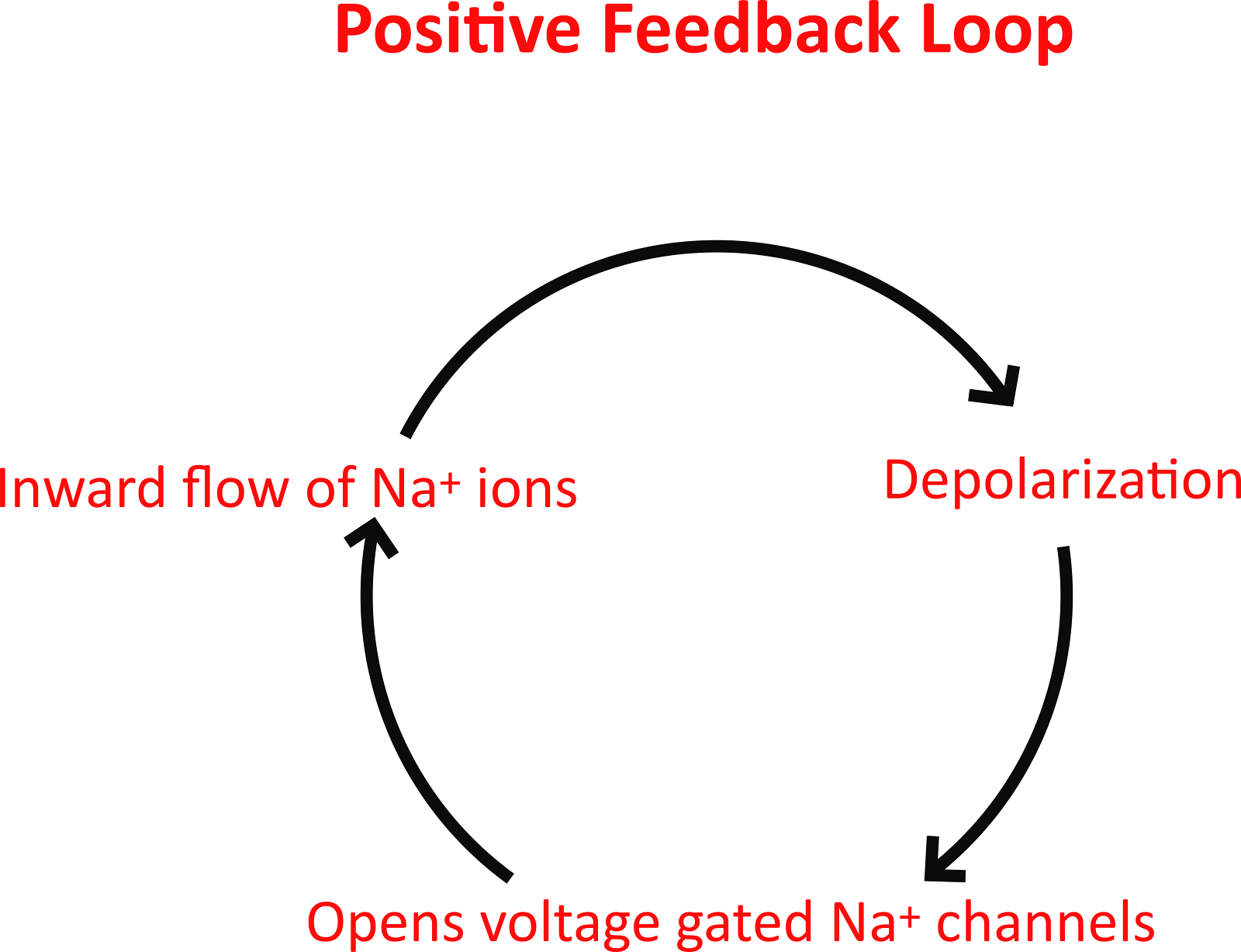
Author: DrV
This increases the membrane potential to around +60 mV. This extreme positivity is termed as overshoot and is the peak of the action potential curve.
The Na+ influx (i.e., the movement of Na+ into the cell) is only for a short time, and the channels then enter an inactive phase. After a few milliseconds, they enter a resting state. They can now be activated again.
Now that the membrane potential has changed, K+ channels begin to open. In contrast to Na+ channels, K+ channel opening is slower and prolonged.
Author: DrV
The movement of K+ ions outwards that follows completes the process of repolarization. The voltage drops to a value lower than -70mV, and this is called hyperpolarization. After this, it stabilizes to pre-stimulus levels.
Refractory Period
Simply put, it is a time when excitability is low. The absolute refractory period is the period from firing to about 1/3rd into repolarization. During this time, no matter how strong the stimulus, you will not have an action potential.
During depolarization, all the Na+ channels are already open and rapidly letting in Na+ ions, so what can a new AP achieve? During the initial stages of repolarization, the Na+ channels are being inactivated and recover. Only after that can a new AP achieve anything.
The relative refractory period is after the initial phases of repolarization when some Na+ channels have recovered. During this time, an AP can be produced, but it requires a stimulus much stronger than the threshold potential.
Conduction of the Action Potential
Try and Imagine a nerve cell at rest. The inside is negative, and the outside is positive. Now, when there is an AP, this is reversed for a short duration. This causes a “sink”-like phenomenon, where the positive charges immediately adjacent to the AP are pulled away.
This generates a new AP in this newly depolarized part of the neuron.
So what can we conclude from this? The AP does not “travel”, but rather triggers a new AP at the next segment of the neuron. Hence, action potentials are propagated and do not travel like electricity.
The Synapse
We briefly discussed the synapse in the introduction. A synapse consists of:
- Presynaptic membrane: This refers to the neuron carrying the message to be passed
- Synaptic cleft: It is the space where the neurotransmitters are poured and taken up
- Postsynaptic membrane: This refers to the neuron receiving the message to be passed.
Presynaptic membranes store vesicles containing neurotransmitters (more on them coming soon). These, in response to a propagated AP, are pushed to the membrane and secreted out into the synaptic cleft. This is via the influx of Ca+ ions, which cause the exit of these neurotransmitters.
Neurotransmitters then bind to ligand-gated channels on the postsynaptic membrane, which cause then lead to a new AP by altering ion channels.
Neurotransmitters
These are like languages to neurons. There are lots of different types of neurotransmitters, but their purpose is communication.
They can be excitatory or inhibitory. Being an interesting topic themselves, neurotransmitters will be covered in detail later on. In short, however, some neurotransmitters are explained below:
Glutamate is the major excitatory neurotransmitter in the CNS. There are 3 types of Glutamate receptors: AMPA, Kainate, and NMDA receptors. There are various subtypes.
GABA (Gamma-amino-butyric-acid) is the main inhibitory neurotransmitter in the CNS. As for glutamate, GABA also has various types of receptors.
Acetylcholine is a rather popular neurotransmitter for its role at the neuromuscular junction, causing muscle contraction. But it has other roles too, such as autonomic ganglia, septal nuclei and nucleus basalis and many more.
Other neurotransmitters include catecholamines (epinephrine, norepinephrine, and dopamine), serotonin, and histamine, to name a few. There are larger molecules which are called neuropeptides. They are being studied extensively and have applications in a range of treatments.
References:
- Ganong’s Review of Medical Physiology 26th Edition - Page no: 87 - 92
- https://www.ncbi.nlm.nih.gov/books/NBK10921/
Author’s footnote
Neuronal conduction and action potential are high yield concepts for various competitive exams. This physiological aspect of the nervous system can make it easier for us to understand the various pathophysiological processes of many neurological diseases.
Feel free to click on the references for a more in-depth reading if you so desire.
If you feel any information can be added or that there are any inadvertent errors, feel free to let us know in the comments below or bring it to our notice via support@medicalwizardry.com.
Ever otherwise, feel free to use the comments section for discussion.
We would love to hear from you. Reach out to us via admin@medicalwizardry.com to share any random gibberish/ideas that you would like implemented.